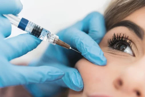
Hyaluronidase is something like a safety net in aesthetic medicine, which gives a piece of mind to all practitioners. Precision (along with safety) is most important in modern aesthetic practice, and nowhere is this more evident than in how we correct and refine dermal filler results.
As hyaluronic acid-based fillers continue to dominate the market, hyaluronidase remains one of the most indispensable tools in a clinician’s arsenal, helping you resolve complications, restoring anatomical balance and patient confidence.
Yet with more advanced filler technologies hitting the market practically on a monthly basis and increasingly diverse patient presentations, the way we use hyaluronidase in 2025 demands a higher level of strategic thinking.
Today, we aim to provide you with the most up-to-date, field-tested hyaluronidase protocols for managing overcorrection and filler migration. Whether you’re fixing a subtle asymmetry or attacking a complex case of delayed migration, the aim is the same: precision and safety.
Hyaluronidase: Mechanism and Indications
Many of you already know, but let’s go over hyaluronidase features first. It is an enzyme that catalyzes the breakdown of hyaluronic acid (HA). Its ability to depolymerize HA makes it uniquely suited for reversing HA-based dermal fillers, either partially or entirely.
The main indications in aesthetics include:
- Overcorrection: When too much filler has been placed, either in volume or in anatomical mismatch.
- Filler migration: When filler moves from its original placement, often seen with superficial perioral injections or in the tear troughs.
- Vascular compromise: An urgent use case where intravascular filler occludes blood flow.
- Aesthetic refinement: Softening edges or asymmetries to perfect a result.
Protocols for Hyaluronidase Use in Overcorrection
When managing overcorrection, the goal is to fix the problem, but also preserve soft tissue integrity.
1. Thorough Assessment First
Begin with an evaluation to determine whether the apparent overcorrection is truly filler-related:
- Palpation remains key. You want to feel for gel consistency versus firm fibrotic tissue or soft edema. Filler generally feels doughy or gel-like, whereas fibrosis presents as firm bands, and edema is fluctuating and mobile.
- Detailed patient history matters. Document product brand, viscosity, rheology, injection technique, and exact timing of previous treatments. Many complications stem from filler layering over several years with incomplete metabolism in between.
- Ultrasound guidance is becoming standard. High-frequency ultrasound (15–20 MHz and above) can visualize retained HA filler, including its depth and distribution.
Keep in mind that not every patient who looks “overdone” is overfilled. Aging changes, fluid retention, or even facial asymmetry can mimic overcorrection.
2. Dosing Recommendations
Gone are the days of large, undifferentiated doses. Here’s how experienced injectors are customizing treatment today:
- 10–30 units per 0.1 mL of HA filler is the general working range. This varies based on the age and density of the product.
- Low, conservative dosing (around 10 units) is preferred for the lips, tear troughs, or fine lines.
- More crosslinked or cohesive products (those using Vycross technology or thick biphasic gels) require higher doses or staged sessions. It’s not uncommon to need 30–75 units per site.
Importantly, each brand and filler type behaves differently under hyaluronidase. A Revance filler will respond differently from a Restylane product, even at the same site.
3. Layered Dissolution: The Gold Standard
High-level clinics now favor a staged approach to correction.
- Inject targeted, fractional doses based on clinical palpation or ultrasound. Spread these over 7–14 days, depending on tissue response and the urgency of correction.
- Reassess between sessions. Volume settles after 24–72 hours, but patient perception often lags behind. Check for unexpected deflation and return of asymmetry.
Minimal trauma and low diffusion are priorities.
Protocols for Managing Filler Migration
Filler migration isn’t always immediately visible, but its effects can drastically impact facial proportions and patient satisfaction.
Knowing where and why migration happens is essential for both prevention and correction:
- Lips: Migration into the white roll, vermillion border, or perioral area is frequently due to superficial injections or high-volume techniques. Lip movement pushes filler upwards over time, particularly with overcorrection.
- Tear troughs: This zone has thin dermis and high mobility. The product here tends to shift medially or inferiorly, especially if the filler is soft or injected superficially. Even a few millimeters of movement can cause a different appearance.
- Cheeks and chin: Filler can track along the SMAS or deeper muscular planes. Migration in these zones is harder to detect.
Poor technique and product selection are the leading culprits. Skin laxity, facial animation, and massage or compression post-treatment can all accelerate unwanted spread.
Precise Dissolution Approach
Migrated filler requires a different strategy than classic overcorrection.
- Use fine-gauge needles or blunt-tip cannulas (27–30G) for precision. Sharp needles are accurate, and cannulas reduce bruising.
- Administer small aliquots (5–15 units) directly into the migration zone. The goal is to target the misplaced product while minimizing enzyme exposure to neighboring tissues.
Give 24-72 hours for full enzymatic activity. Swelling or irregularity immediately after injection is common and not an indication of failure.
Why Filler Overcorrection and Migration Are More Common Than Ever?
Aesthetic trends have matured. Patients are no longer requesting the exaggerated looks that once defined the early filler era. Instead, they’re asking for subtler changes.
This shift is revealing problems in prior treatments:
- Excess product layered over time, especially in midface and periorbital zones: Many patients have received repeat treatments without full reassessment of existing filler, leading to unintended volume buildup. This is especially noticeable in delicate areas like the tear trough, where even small amounts of residual filler can distort natural contours.
- Low-viscosity fillers migrating into surrounding tissues: Softer gels, particularly those used for superficial correction, can spread under muscle movement or with time. This often results in puffiness or unnatural fullness in areas like the nasolabial fold or lateral cheeks, even if the injection site was more centralized.
- Delayed swelling or nodules, sometimes misread as product or edema: What appears to be persistent swelling may actually be late-onset inflammatory response or encapsulated filler. Without ultrasound or experienced palpation, these issues can go unnoticed and lead to unnecessary additional filler.
- Poor technique or superficial placement, especially in high-mobility areas: Injections placed too close to the surface or in the wrong plane can quickly become visible or migrate. This is common in lips, marionette lines, and even along the jawline, where muscle movement significantly influences filler behavior.
Preventing Overcorrection and Migration: Best Practices in 2025
When prevention is part of your protocol, you may not need hyaluronidase.
- Ultrasound mapping: Now a routine tool in many clinics, helps visualize anatomy and previous filler.
- Lower volumes per session: Especially in the midface and lips.
- Product selection: Choosing a filler with rheological properties appropriate for the site (e.g., avoiding overly soft gels in mobile areas).
- Education: Set realistic expectations with patients. Less is often more and touch-ups are preferable to reversal.
Final Thoughts
In day-to-day practice, hyaluronidase has become part of how we plan, adjust, and sometimes completely rethink treatment goals/results. Especially in cases of overcorrection or migration, the decision to use it requires clinical judgment, experience, and a good understanding of how filler behaves over time.
By 2025, the time when most HA treatments were very similar is long gone. Nowadays, you can fix superficial Tyndall effect, delayed migration, or a patient who simply doesn’t feel like themselves after a treatment elsewhere. But the key is approaching each case methodically. We assess. We wait when needed. We re-treat carefully, and only when the tissue tells us it’s ready.
More than anything, the conversation around hyaluronidase is changing. It’s less about “fixing mistakes” and more about optimizing results. Done right, it doesn’t undo your work. It enhances your reputation for getting it right.
Frequently Asked Questions (FAQs)
How should hyaluronidase protocols be adapted for patients with autoimmune conditions?
In autoimmune patients, a more conservative dose and careful allergy history are a must. Pre-treatment with antihistamines or low-dose steroids may reduce the risk of flares. Always conduct a thorough risk-benefit analysis before doing it.
Can hyaluronidase be used to prevent filler migration in high-risk anatomical zones?
Hyaluronidase is not used preventively, but early intervention at the first signs of migration can limit its extent. Zones like the tear trough or perioral area are exceptionally prone to movement, so product selection and precise placement matter more.
How does hyaluronidase interact with newer hybrid fillers that contain both HA and calcium hydroxylapatite?
Hyaluronidase will only target the HA component, leaving calcium hydroxylapatite (CaHA) intact. This can lead to partial resolution but not full reversal. Practitioners need to be cautious and manage patient expectations accordingly.
What are the best practices for combining hyaluronidase with ultrasound-guided injections?
Ultrasound helps visualize filler deposits, vascular structures, and target tissue planes, improving precision. It’s particularly valuable in complex corrections or deeply placed products. Practitioners trained in sono-guided injection see fewer complications.
References
Murray G, Convery C, Walker L, Davies E. Guideline for the Safe Use of Hyaluronidase in Aesthetic Medicine, Including Modified High-dose Protocol. J Clin Aesthet Dermatol. 2021 Aug;14(8):E69-E75. Epub 2021 Aug 1. PMID: 34840662; PMCID: PMC8570661.
Jung H. Hyaluronidase: An overview of its properties, applications, and side effects. Arch Plast Surg. 2020 Jul;47(4):297-300. doi: 10.5999/aps.2020.00752. Epub 2020 Jul 15. PMID: 32718106; PMCID: PMC7398804.
Murray G, Convery C, Walker L, Davies E. Guideline for the Management of Hyaluronic Acid Filler-induced Vascular Occlusion. J Clin Aesthet Dermatol. 2021 May;14(5):E61-E69. Epub 2021 May 1. PMID: 34188752; PMCID: PMC8211329.
DeLorenzi C. New High Dose Pulsed Hyaluronidase Protocol for Hyaluronic Acid Filler Vascular Adverse Events. Aesthet Surg J. 2017 Jul 1;37(7):814-825. doi: 10.1093/asj/sjw251. PMID: 28333326.
King M. Management of Tyndall Effect. J Clin Aesthet Dermatol. 2016 Nov;9(11):E6-E8. Epub 2016 Nov 1. PMID: 28210392; PMCID: PMC5300720.


0 Comments